Organization of treatment and rehabilitation in Germany.
Complete medical support.
Complete medical support.
+49 176 17917001
All departments

Melanoma is skin cancer that develops from the skin's pigment cells (melanocytes). In Russia, it accounts for 1-3% of all cancer cases and occurs even in children. More than 70% of disease cases arise at the site of a benign mole — a pigmented nevus that malignizes under the influence of unfavorable endogenous and/or exogenous factors.
Melanoma is considered the most aggressive tumor in oncology; it tends to grow rapidly and metastasize, which significantly worsens the prognosis for the patient. In this regard, early diagnosis and treatment of melanoma in Germany is particularly relevant: when therapy begins at the first stage, when the tumor is located only within the epidermis, the probability of cure approaches 100%.
Important! Germany has achieved one of the lowest mortality rates from melanoma — about 1.3%.The prognosis for 5-year survival with tumor thickness up to 1 mm and no metastases is more than 90%, and with penetration depth of more than 4 mm without metastasis — up to 50%. In case of distant metastases, 5-year survival does not exceed 15-20%.
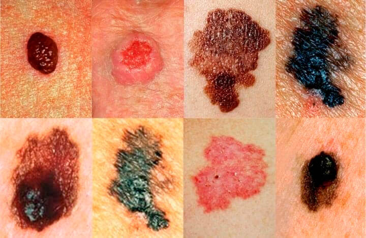
Melanoma is characterized by changes in the size and structure of a previously existing pigmented nevus, or the appearance of an "unusual" mole that differs from others in color, shape, and surface character. The tumor appears as a round, triangular or polygonal formation of dark brown, gray, black or bluish color. The coloring is often uneven, spotted. Standard sizes range from a few millimeters to 2-3 cm.
On the surface of the pigmented neoplasm, ulceration, weeping, bleeding often occur, and healing of skin lesions proceeds with the formation of crusts. In 20% of cases, melanoma has a nodular form: it rises above the skin in the form of a mushroom-shaped formation.
Melanoma most often metastasizes to the skin and lymph nodes, and distant metastases to the liver, kidneys, brain, lungs and other internal organs are also possible.

Each stage has clinical subtypes, which are designated by Latin letters A, B, C. The type and stage of the tumor can be determined most accurately after an extended examination of the patient.

The examination begins with a standard physical examination of the suspicious mole, collection of complaints and disease history. To quickly identify suspicious neoplasms, dermato-oncologists use the ABCDE algorithm, which takes into account asymmetry, contours, color, diameter and dynamics of tumor size changes. The following research methods are prescribed for diagnosis at the clinic:
Special attention is paid to melanoma diagnostics in Germany, since the success of treatment largely depends on the timeliness and reliability of the diagnosis. Many patients from CIS countries come to German clinics to undergo examination with unique equipment and receive an expert medical opinion in a short time.

Maximum possible excision of the tumor within healthy tissues is the optimal method of melanoma treatment in Germany, which shows maximum effectiveness at an early stage of cancer. With timely surgical intervention, it is possible to completely remove malignant cells to prevent disease recurrence. However, most patients see a doctor when melanoma is already progressing and metastasizing, so radical operations are not always possible.
For effective melanoma removal, doctors make wide incisions capturing 2-3 cm of unchanged skin — this way it is possible to reduce the risk of tumor recurrence in the same location. After the primary operation, cosmetic defects remain, for the elimination of which autoplasty methods with the patient's own skin flap are provided.
In German clinics, as an alternative to classical surgical treatment, modern minimally invasive methods can be used: cryodestruction of the neoplasm at an early stage, stereotactic radiosurgery for targeted and bloodless tumor excision, laser removal of malignant cells.
Classical treatment protocols prescribe immunochemotherapy with alpha-interferon drugs in adjuvant mode (after tumor removal). Chemotherapy shows the best results in patients with second and third stages of melanoma, while in the first stage treatment is possible without medication, and in the fourth — the effectiveness of adjuvant therapy has not been proven.

Melanoma treatment in Germany shows good results due to the combination of traditional and modern therapy directions. German doctors treat skin cancer using the best achievements of world medicine, and are engaged in the development and implementation of proprietary treatment methods.
The following directions are used in comprehensive melanoma therapy programs at German clinics:
In addition, experimental treatment is available in some German clinics that has not yet been performed in any country in the world. Patients have a chance to participate in clinical trials of the latest drugs to receive innovative treatment for free and get a chance for success even at the terminal stage of melanoma. German doctors were among the first to develop and test in practice a unique vaccine against melanoma that changes the function of the patient's immune system. So far, this drug is only available within clinical trials.
The cost of the full course of diagnosis and therapy is determined individually for each patient after analysis of medical documentation by the receiving German clinic. The final amount depends on the volume of additional studies, the necessity and method of surgical operation, and the possibility of innovative methods.
Approximate prices for melanoma diagnostics in Germany start from €530, and the cost of histological examination of tumor biomaterial can reach €3000. Radical tumor removal costs from €3160, targeted treatment from €1700, and the price of modern immunotherapy directions starts from €20000.
Melanoma treatment is carried out at the following German clinics:
as well as Nuremberg Clinic, University Hospital Freiburg, Asklepios Clinic, University Hospital Cologne.
Timely contact is one of the main factors for successful melanoma treatment. Leave a request in the online form on this page, and our medical consultant will contact you shortly to select a clinic and discuss all the details of treatment in Germany.

High-precision research methods

Comprehensive approach to treatment

New surgical techniques

Low complication rate

Treatment of complex cases

High therapy effectiveness
High-precision research methods
Comprehensive approach to treatment
New surgical techniques
Low complication rate
Treatment of complex cases
High therapy effectiveness
 +49 176 17917001
Or send us a message:


