
Organization of treatment and rehabilitation in Germany.
Complete medical support.
Complete medical support.


+49 176 17917001
All departments

Left image – before surgery: cervical disc herniation C5/C6 protruding 5 mm into the spinal canal.
Right image – 6 weeks after surgery: the disc herniation is no longer visible.

Left image – before surgery: complete traumatic shoulder dislocation, bony Bankart lesion, extensive labral damage, rupture of the superior glenohumeral ligament, partial tears of the supraspinatus and infraspinatus tendons, tear at the base of the biceps tendon.
Right image – 5 years after surgery: regenerative reconstruction of the shoulder joint; the Hill-Sachs defect and the bony Bankart lesion are no longer visible.

Left image – before surgery: grade 4–5 degeneration of the lumbar disc L5/S1 – the disc is almost absent, with bone contact between the vertebrae.
Right image – 12 weeks after surgery: restoration of disc height, volume and shape after transplantation of an autologous regenerative concentrate into the L5/S1 disc.

Left image – before surgery: grade 4 osteoarthritis, osteonecrosis and meniscal degeneration of the left knee joint.
Right image – after surgery: regeneration of the tissues in the areas of osteonecrosis, osteoarthritis and meniscal degeneration.

Left image – before surgery: osteoarthritis of the atlanto-occipital joint and the median atlanto-axial joint.
Right image – 5 months after surgery: restoration of the atlanto-occipital and median atlanto-axial joints with complete restoration of the joint cartilage; signs of osteoarthritis are no longer detectable.

Left image – before surgery: acute traumatic right-sided mediolateral herniation of 8 mm at C7/Th1, spinal canal stenosis, absolute stenosis of the right C7/Th1 foramen, spinal cord compression with displacement to the left and damage to the C8 nerve root.
Right image – 2 months after surgery: marked reduction of the herniation, elimination of spinal cord compression and displacement, sharp reduction of the foraminal stenosis with decompression of the right C7/Th1 root.

Left image – before surgery: right-sided mediolateral sequestered disc herniation L5/S1, 11 mm, causing stenosis of the spinal canal and the intervertebral foramen with compression of the L5 and S1 nerve roots.
Right image – 14 weeks after surgery: the sequestered fragment is no longer visible; the spinal canal and the right intervertebral foramen show no stenosis, with complete decompression of the L5 and S1 nerve roots.
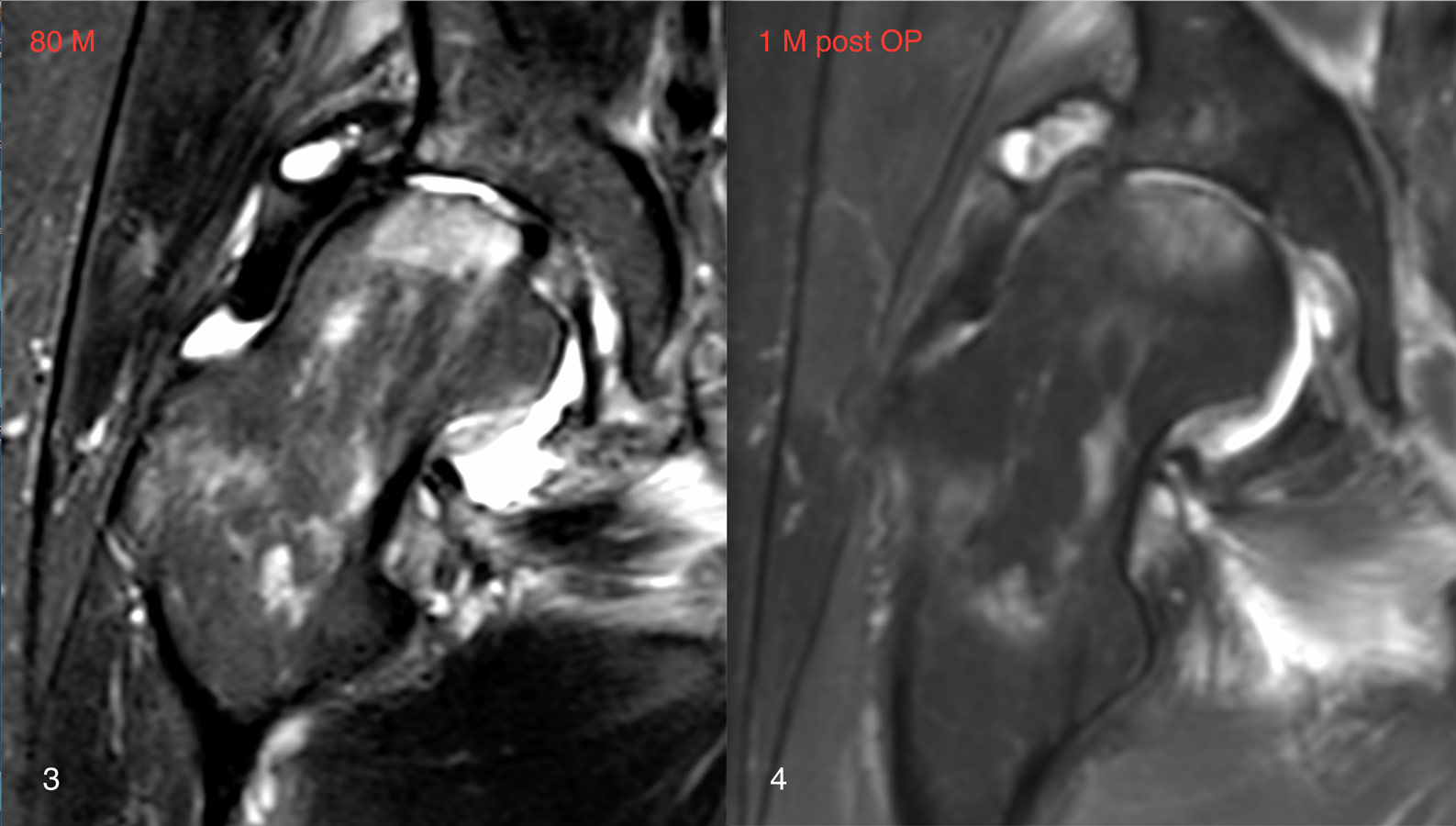
Left image – before surgery: grade 4 hip osteoarthritis, osteonecrosis of the femoral head and neck, synovitis.
Right image – 1 month after surgery: restoration of the joint surfaces; osteoarthritis and synovitis are not visible; signs of intensive bone regeneration in the areas of necrosis.

Left image – before surgery: osteonecrosis of the lateral condyle of the right knee joint.
Right image – 6 months after surgery: complete restoration of the bone tissue in the area of osteonecrosis; signs of osteonecrosis are no longer visible.
For everyone planning to undergo treatment in Germany, reviews are an important criterion for selecting a reliable medical tourism company. Doktor Munchen is happy to share with you some reviews about treatment in German clinics sent by our patients. We are very pleased that we have already been able to help many people. Come for diagnostics and treatment in Germany!
To send your review, please use the contact form.
In your review, describe your condition before arrival, which clinic you underwent diagnostics and/or treatment at in Germany, how you feel after treatment. And don't forget to indicate your age. We do not publish the surnames of patients who left reviews.

Parkinson's disease and movement disorders

Neurosurgery, Gamma Knife

Gynecology

Orthopedics, Hip surgery

Surgical treatment

Orthopedics, Knee surgery


