Fracture Nonunion (Pseudoarthrosis) Treatment in Germany
Fracture nonunion (pseudoarthrosis, false joint, Pseudoarthrose) is a condition in which the bone fails to heal within the expected timeframe and the consolidation process ceases. Fibrous or cartilaginous tissue forms at the fracture site instead of bone, creating a "false joint" with pathological mobility. Nonunion develops in 5–10% of all fractures and is one of the most challenging problems in traumatology. German clinics employ modern methods of bone healing stimulation: autologous bone grafting, BMP, Ilizarov apparatus, and innovative MIBRAR® therapy with autologous stem cells.
What is Fracture Nonunion
Normally, a fracture heals in 3 stages: inflammation (1–2 weeks), callus formation (4–12 weeks), remodeling (months–years). If 6–9 months after fracture no radiographic progression of healing is observed, nonunion is diagnosed. Delayed union is diagnosed when healing has not occurred within the usual timeframe, but the process continues.
Pseudoarthrosis forms when the fragment ends become covered with fibrous tissue or cartilage, and a gap resembling a joint space develops between them. With prolonged existence, a synovial membrane and capsule form — a "true" false joint.
Types of Pseudoarthrosis
Type
X-ray Findings
Biology
Treatment
Hypertrophic ("elephant foot")
Widened, sclerotic ends with abundant callus
Adequate blood supply, insufficient stability
Stable osteosynthesis (without bone grafting)
Atrophic
Thinned, rounded ends without callus
Insufficient blood supply + instability
Stabilization + bone grafting + biostimulation
Oligotrophic
Minimal callus, gap
Adequate blood supply but insufficient stimulation
Decortication + stabilization
Infected
Sequestra, foci of destruction, fistulae
Infection + disrupted blood supply
Debridement + antibiotics + staged reconstruction
Causes
Insufficient fixation — unstable osteosynthesis, poor reduction, early hardware removal. Movement at the fracture site → fibrosis instead of bone.
Disrupted blood supply — femoral neck, scaphoid, and talus fractures — areas with poor blood supply. Extensive soft tissue trauma.
Bone defect — loss of a bone fragment in an open fracture or surgery. Gap > 1 cm.
Infection — postoperative infection destroys regenerating bone tissue. Osteomyelitis.
Systemic factors — osteoporosis, diabetes mellitus, smoking (reduces blood flow by 25%), NSAID and corticosteroid use, vitamin D deficiency, poor nutrition.
High-energy trauma — open fractures, multiple fractures, gunshot fractures.
Symptoms
Weight-bearing pain — persistent or recurrent pain at the fracture site 3–6 months after injury.
Pathological mobility — sensation of movement at the fracture site under load or palpation.
Functional impairment — inability to bear full weight, limping, restricted motion in adjacent joints.
Limb deformity — angular or rotational deviation, shortening.
Swelling — persistent soft tissue swelling at the fracture site.
In infected pseudoarthrosis: fistulae with purulent discharge, skin redness, general malaise.
Electrical stimulation — electromagnetic fields stimulate osteoblasts. Used for superficial pseudoarthrosis.
Systemic factor correction — smoking cessation (!), vitamin D and calcium normalization, diabetes control, NSAID discontinuation.
Brace stabilization — for stable hypertrophic pseudoarthrosis combined with ESWT/LIPUS.
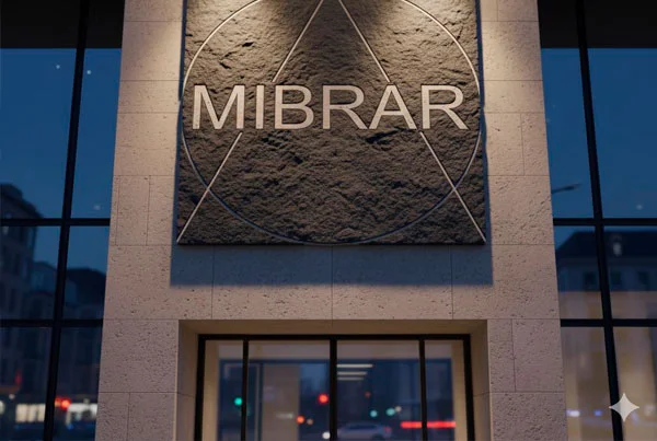
MIBRAR® Therapy
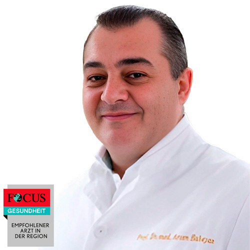
Fracture nonunion (Unheilbarkeit der Frakturen, Pseudoarthrose) is included in the list of indications for MIBRAR® technology. Professor Babayan's method offers microinvasive stimulation of bone regeneration with autologous material.
MIBRAR® for fracture nonunion:
ARK transplantation to the fracture zone — autologous regenerative concentrate contains mesenchymal stem cells (CD34+ from blood + lipogenic from subcutaneous fat) that differentiate into osteoblasts and form new bone tissue.
MIBRAR® microperforations — targeted micro-injuries to sclerotic fragment ends open channels for stem cell migration and vessel ingrowth. Mimics the decortication principle without open surgery.
Growth factors — BMP (bone morphogenetic proteins) and VEGF (vascular endothelial growth factor) in ARK stimulate osteogenesis and angiogenesis.
Sono Control Arm™ — concentrate delivery accuracy of 0.1 mm directly into the fracture gap, without X-ray radiation.
The procedure is outpatient, without anesthesia. Can be used independently or as a supplement to surgical stabilization. Especially effective in atrophic pseudoarthrosis where the key problem is insufficient biological stimulation.
Surgical Treatment
Re-osteosynthesis with autologous bone grafting — removal of non-viable tissue, stable fixation with plate or nail + autograft from the iliac crest. Gold standard for atrophic pseudoarthrosis.
Judet decortication — removal of the cortical plate around the fracture zone to stimulate blood supply. For hypertrophic type + stable fixation.
Compression osteosynthesis (Ilizarov apparatus) — external fixator with gradual compression of the fracture zone. Indicated for infected pseudoarthrosis, bone defects. Maslov–Ilizarov method: bone segment transport to close the defect.
BMP (bone morphogenetic protein) — BMP-2 (InFuse), BMP-7 (OP-1). Powerful osteogenesis stimulation. Used for severe atrophic pseudoarthrosis.
Vascularized bone graft — transplantation of a bone segment with a vascular pedicle (fibula). For large defects and disrupted blood supply.
Masquelet technique (induced membrane) — two-stage: (1) cement spacer placement → induced membrane formation; (2) after 6–8 weeks — cement removal + bone grafting inside the membrane. For defects > 4 cm.
Professor Babayan's specialized center. Treatment of spine and joint diseases using the patented MIBRAR® technology — no incisions, no anesthesia, outpatient. More than 25,000 successful procedures. The world's only center offering the full range of MIBRAR® techniques.
13 специализированных лечебных центров и 3 амбулатории. Основное отделение работает в Верхней Баварии и находится в одном из самых живописных мест на берегу озера Тегернзее.
After Berlin's Charité, the Munich University Hospital with the Innenstadt and Großhadern campus is the largest maximum care medical complex in Germany.
The Department of Hematology and Oncology offers a full range of diagnostic and therapeutic services in these fields. The highly qualified team of doctors provides patients with effective treatment of all oncological diseases, blood and lymph pathologies (e.g., leukemia, multiple myeloma).
A network of multidisciplinary clinics located in five districts of Munich. They provide a high-class range of medical services. The municipal clinics are academic clinics of both Munich universities.
The main advantages of OrthoLiga clinics are: highly qualified specialists, world-class medical care, state-of-the-art diagnostics, and comprehensive patient care.
Advantages of Fracture Nonunion Treatment in Germany
After treatment ends, within 10 days you receive the final invoice and copies of invoices from the clinic. The remaining amount is returned to the card within 3 days.
After treatment ends, within 10 days you receive the final invoice and copies of invoices from clinics. The remaining amount is returned to the card within 3 days.


 +49 176 17917001