Perthes disease (Legg–Calvé–Perthes disease, Morbus Perthes) is avascular necrosis of the ossification center of the femoral head in children. The disease develops due to disrupted blood supply to the femoral epiphysis, leading to femoral head deformation and early coxarthrosis. It most commonly affects boys aged 4–8 years (M:F ratio = 4:1). Bilateral involvement occurs in 10–15% of cases. German clinics employ precise staging methods, containment therapy, and innovative MIBRAR® therapy to stimulate bone tissue regeneration.
What is Perthes Disease
In Perthes disease, blood supply to the epiphysis (head) of the femur is disrupted. The epiphysis is the bony nucleus that in children is still in the process of growth and formation. Without adequate blood flow, the bone tissue of the epiphysis dies (necrosis), then gradually resorbs and is replaced by new bone (reossification). The entire cycle takes 2–4 years. The main problem is that during the fragmentation phase, the softened femoral head deforms under load. If the head loses its spherical shape, joint incongruence develops → early arthrosis in adulthood.
The goal of treatment is to preserve the spherical shape of the femoral head by ensuring its containment within the acetabulum throughout the entire disease cycle.
Disease Stages
Stage
Name
X-ray Findings
Duration
I
Initial (necrosis)
Epiphyseal condensation, joint space widening
6 months
II
Fragmentation
Epiphyseal breakdown into fragments, areas of lucency and sclerosis
8–12 months
III
Reossification
Restoration of bone structure, new bone formation
12–24 months
IV
Outcome (remodeling)
Final head shape — from spherical to "mushroom-shaped"
Until growth completion
Critical phase — stage II (fragmentation). It is during this period that the softened head is susceptible to deformation. All treatment efforts are directed at preserving the head shape during this phase.
Causes and Risk Factors
Disrupted epiphyseal blood supply — the exact cause is unknown. Thrombosis or spasm of the vessels supplying the femoral head (medial circumflex artery) is suspected.
Heredity — familial predisposition in 5–10% of cases.
Constitutional features — children with Perthes disease are often shorter and thinner than their peers. Delayed bone age.
Parental smoking — passive smoking is associated with increased risk (microcirculation impairment).
Hyperactivity and injuries — repeated microtraumas of the hip joint.
Thrombophilia — congenital coagulation disorders (protein C and S deficiency, Factor V Leiden mutation).
Symptoms
Limping — the main symptom! Painless or mildly painful limping in a child aged 4–8 years is a reason for examination.
Groin or knee pain — hip joint pain often radiates to the knee. "Knee pain" in a child — always check the hip joint!
Restricted movement — decreased abduction and internal rotation of the hip. Adductor muscle spasm.
Fatigue while walking — the child asks to be carried, refuses long walks.
Leg length discrepancy — with femoral head deformation and femoral neck "collapse".
Thigh and gluteal muscle atrophy — on the affected side.
Important: the disease develops gradually, without fever or signs of infection. Limping lasting > 2 weeks in a child is an absolute indication for hip X-ray.
Clinical examination — gait assessment (Trendelenburg limp), range of motion (decreased abduction and rotation), limb length, muscle atrophy.
Hip X-ray — AP and Lauenstein views. Waldenström staging. Assessment of head shape, epiphyseal height, Shenton line.
MRI — early diagnosis (before radiographic changes). Visualization of the necrosis zone, viable tissue, cartilage condition. "Head at risk" assessment.
Ultrasound — quick method for detecting hip joint effusion (transient synovitis as a differential diagnosis).
Arthrography — contrast study of cartilaginous head shape (wider than bony). Performed before surgery for containment planning.
Laboratory tests — ESR, CRP, leukocytes — to rule out septic coxitis and juvenile arthritis.
Classifications
Herring classification (lateral pillar): assesses the height of the lateral pillar of the epiphysis during the fragmentation phase. The most prognostically significant:
Group A — lateral pillar not affected. Good prognosis.
Group B — lateral pillar height > 50%. Moderate prognosis.
Group B/C — borderline. Requires careful monitoring.
Group C — lateral pillar height < 50%. Poor prognosis without treatment.
Catterall classification: assesses the extent of head involvement (4 groups). Catterall risk factors ("head at risk"): lateral calcification, horizontalization of the growth plate, subluxation, metaphyseal cysts.
Conservative Treatment
Indicated with favorable factors: age < 6 years, Herring group A or B, small extent of involvement:
Observation — regular radiographic monitoring every 3–4 months. In Herring group A children < 6 years — good prognosis without active treatment.
Containment bracing — abduction braces (Atlanta brace, Scottish Rite brace) keep the femoral head within the acetabulum, protecting it from deformation. Worn for 12–18 months.
Physical therapy — exercises to maintain range of motion, adductor stretching, strengthening of abductor muscles.
NSAIDs — for pain management.
Traction — for severe muscle spasm and subluxation.
MIBRAR® Therapy
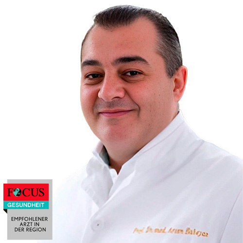
Perthes disease (Morbus Perthes) is included in the list of indications for MIBRAR® technology (individual assessment). Professor Babayan's method offers a fundamentally new approach — stimulating epiphyseal bone tissue regeneration with autologous material.
MIBRAR® for Perthes disease:
Revascularization stimulation — ARK (autologous regenerative concentrate) growth factors stimulate angiogenesis — restoration of blood supply to the necrosis zone.
Accelerated reossification — mesenchymal stem cells differentiate into osteoblasts, accelerating replacement of necrotic bone with new bone.
Head shape preservation — faster reossification reduces the period during which the softened head is susceptible to deformation.
Microinvasiveness — Sono Control Arm™ ensures precise ARK delivery to the necrosis zone through a puncture (without incision), which is critically important for a child's growing bone.
The procedure is outpatient, without general anesthesia (sedation is possible). Only autologous material is used — full biocompatibility, no side effects.
Surgical Treatment
Surgery is indicated with unfavorable factors: age > 8 years, Herring group B/C or C, "head at risk", subluxation, loss of containment:
Varus intertrochanteric femoral osteotomy — tilting the femoral neck, "deepening" the head into the acetabulum. The most common operation for Perthes disease.
Pelvic osteotomy (Salter, Triple) — rotation of the acetabulum for better head coverage. Indicated for lateral coverage deficiency.
Combined (femur + pelvis) — for severe cases with bilateral deficiency.
Arthrodiastasis — application of an external fixator with joint distraction. Unloads the head during the fragmentation period.
Shelf procedure — a bony "shelf" over the femoral head for additional coverage.
Professor Babayan's specialized center. Treatment of spine and joint diseases using the patented MIBRAR® technology — no incisions, no anesthesia, outpatient. More than 25,000 successful procedures. The world's only center offering the full range of MIBRAR® techniques.
13 специализированных лечебных центров и 3 амбулатории. Основное отделение работает в Верхней Баварии и находится в одном из самых живописных мест на берегу озера Тегернзее.
After Berlin's Charité, the Munich University Hospital with the Innenstadt and Großhadern campus is the largest maximum care medical complex in Germany.
The Department of Hematology and Oncology offers a full range of diagnostic and therapeutic services in these fields. The highly qualified team of doctors provides patients with effective treatment of all oncological diseases, blood and lymph pathologies (e.g., leukemia, multiple myeloma).
A network of multidisciplinary clinics located in five districts of Munich. They provide a high-class range of medical services. The municipal clinics are academic clinics of both Munich universities.
This is a large multidisciplinary clinic in Munich offering a wide range of therapeutic services in various areas of pediatrics and related fields of medicine.
The main advantages of OrthoLiga clinics are: highly qualified specialists, world-class medical care, state-of-the-art diagnostics, and comprehensive patient care.
Advantages of Perthes Disease Treatment in Germany
After treatment ends, within 10 days you receive the final invoice and copies of invoices from the clinic. The remaining amount is returned to the card within 3 days.
After treatment ends, within 10 days you receive the final invoice and copies of invoices from clinics. The remaining amount is returned to the card within 3 days.


 +49 176 17917001